Medicine and HealthAnesthesiology
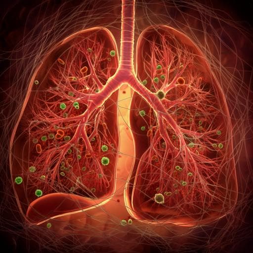
Mechanical Ventilation Induces Neutrophil Extracellular Trap Formation
C. Yildiz, N. Palaniyar, et al.
In a groundbreaking study, researchers including Christopher Yildiz and Nades Palaniyar explored ventilator-induced lung injury (VILI) and uncovered the role of neutrophil extracellular traps (NETs) in this condition. Their findings reveal that high tidal volume mechanical ventilation is essential for NET production, suggesting new avenues for addressing lung injury in clinical settings.
Related Publications
Explore these studies to deepen your understanding
Adjacent work that informs or extends this paper's methodology and findings.

Medicine and Health
Neutrophil Extracellular Trap Formation: Physiology, Pathology, and Pharmacology
M. Ravindran, M. A. Khan, et al.

Medicine and Health
Progression of Cystic Fibrosis Lung Disease from Childhood to Adulthood: Neutrophils, Neutrophil Extracellular Trap (NET) Formation, and NET Degradation
M. A. Khan, Z. S. Ali, et al.
Medicine and Health
Neutrophil Extracellular Trap Formation: Physiology, Pathology, and Pharmacology
M. Ravindran, M. A. Khan, et al.

Medicine and Health
Alkaline pH Promotes NADPH Oxidase-Independent Neutrophil Extracellular Trap Formation: A Matter of Mitochondrial Reactive Oxygen Species Generation and Citrullination and Cleavage of Histone
C. N. D. Souza, L. C. D. Breda, et al.


