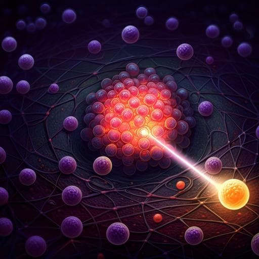
Intelligent drugs based on notch protein remodeling: a defensive targeting strategy for tumor therapy
Y. Sun, Y. Lu, et al.
Explore these studies to deepen your understanding
Adjacent work that informs or extends this paper's methodology and findings.
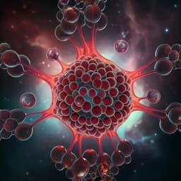
Generation of tumor spheroids using a droplet-based microfluidic device for photothermal therapy
J. M. Lee, J. W. Choi, et al.

Effectiveness of app-based cognitive behavioral therapy for insomnia on preventing major depressive disorder in youth with insomnia and subclinical depression: A randomized clinical trial
S. Chen, J. Que, et al.

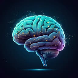
Neuroimaging the effects of smartphone (over-)use on brain function and structure—a review on the current state of MRI-based findings and a roadmap for future research
C. Montag and B. Becker
Neuroimaging the effects of smartphone (over-)use on brain function and structure-a review on the current state of MRI-based findings and a roadmap for future research
C. Montag and B. Becker


