ChemistryNature Communications
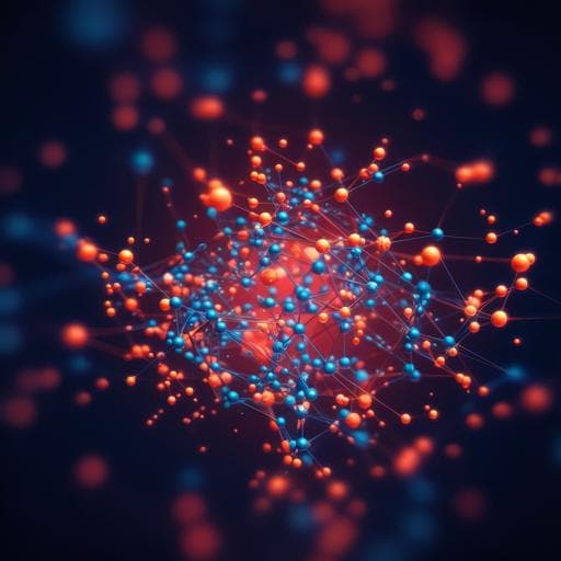
Imaging 3D chemistry at 1 nm resolution with fused multi-modal electron tomography
J. Schwartz, Z. W. Di, et al.
Discover how a team of researchers led by Jonathan Schwartz has tackled the challenge of mapping chemistry in nanoscale materials. Their groundbreaking work introduces fused multi-modal electron tomography, achieving unprecedented high-resolution 3D chemical mapping. This innovative technique promises to revolutionize our understanding of various nanomaterials by providing sub-nanometer resolution even at low electron doses.
Related Publications
Explore these studies to deepen your understanding
Adjacent work that informs or extends this paper's methodology and findings.

Engineering and Technology
Imaging atomic-scale chemistry from fused multi-modal electron microscopy
J. Schwartz, Z. W. Di, et al.
Medicine and Health
Rapid 3D imaging at cellular resolution for digital cytopathology with a multi-camera array scanner (MCAS)
K. Kim, A. Chaware, et al.
Physics
Mixed-state electron ptychography enables sub-angstrom resolution imaging with picometer precision at low dose
Z. Chen, M. Odstrcil, et al.
Biology
In vivo volumetric imaging of calcium and glutamate activity at synapses with high spatiotemporal resolution
W. Chen, R. G. Natan, et al.


