
Medicine and HealthNature Communications
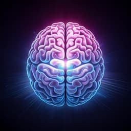
Identification of four biotypes in temporal lobe epilepsy via machine learning on brain images
Y. Jiang, W. Li, et al.
This groundbreaking research, conducted by Yuchao Jiang and colleagues, unveiled four distinct subtypes of temporal lobe epilepsy (TLE) by analyzing MRI data. The study's findings highlight diverse patterns of brain atrophy and their impact on disease progression and treatment outcomes, paving the way for personalized medicine approaches in TLE management.
Related Publications
Explore these studies to deepen your understanding
Adjacent work that informs or extends this paper's methodology and findings.
Medicine and Health
Machine learning explains response variability of deep brain stimulation on Parkinson's disease quality of life
E. Ferrea, F. Negahbani, et al.
Chemistry
Unraveling the energetic significance of chemical events in enzyme catalysis via machine-learning based regression approach
Z. Song, H. Zhou, et al.

Humanities
From remote sensing and machine learning to the history of the Silk Road: large scale material identification on wall paintings
S. Kogou, G. Shahtahmassebi, et al.

Medicine and Health
Machine learning for accurate estimation of fetal gestational age based on ultrasound images
L. H. Lee, E. Bradburn, et al.


