Medicine and HealthCommunications Biology
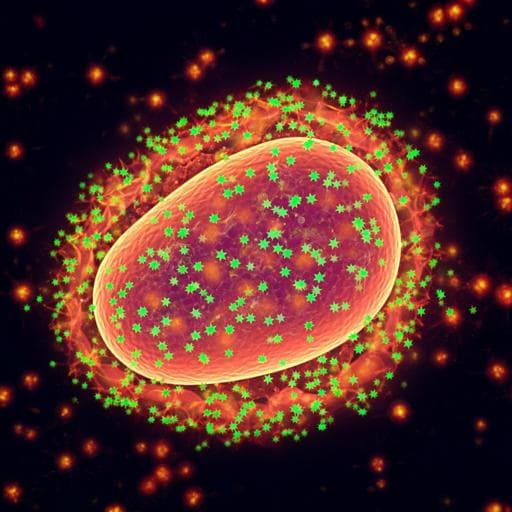
Hyperactive Natural Killer cells in Rag2 knockout mice inhibit the development of acute myeloid leukemia
E. Sugimoto, J. Li, et al.
This groundbreaking study unveils the pivotal role of hyperactive natural killer (NK) cells in combating acute myeloid leukemia (AML). Conducted by a team of researchers, it demonstrates how NK cells can significantly hinder AML progression, especially during tumor evolution. Discover the exciting implications of these findings!
Related Publications
Explore these studies to deepen your understanding
Adjacent work that informs or extends this paper's methodology and findings.

Medicine and Health
Comprehensive characterization of IFNy signaling in acute myeloid leukemia reveals prognostic and therapeutic strategies
B. Wang, P. K. Reville, et al.
Medicine and Health
Environmental signals rather than layered ontogeny imprint the function of type 2 conventional dendritic cells in young and adult mice
N. E. Papaioannou, N. Salei, et al.

Medicine and Health
Off-the-shelf CAR natural killer cells secreting IL-15 target spike in treating COVID-19
T. Lu, R. Ma, et al.

Medicine and Health
Longitudinal single-cell profiling of chemotherapy response in acute myeloid leukemia
M. M. Naldini, G. Casirati, et al.


