Medicine and HealthMucosal Immunology
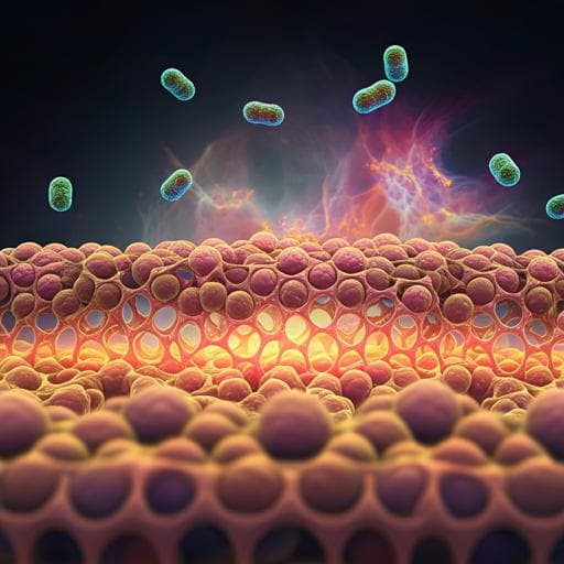
Gut-derived short-chain fatty acids modulate skin barrier integrity by promoting keratinocyte metabolism and differentiation
A. Trompette, J. Pernot, et al.
Discover how a fiber-rich diet can alleviate atopic dermatitis (AD) by strengthening skin barrier functions, thanks to gut-derived short-chain fatty acids like butyrate. This groundbreaking research conducted by Aurélien Trompette, Julie Pernot, and others reveals exciting new insights into skin health.
Related Publications
Explore these studies to deepen your understanding
Adjacent work that informs or extends this paper's methodology and findings.

Medicine and Health
Microbial short-chain fatty acids modulate CD8+ T cell responses and improve adoptive immunotherapy for cancer
M. Luu, Z. Riester, et al.

Medicine and Health
Gut microbiota link dietary fiber intake and short-chain fatty acid metabolism with eating behavior
E. Medawar, S. Haange, et al.

Medicine and Health
The interplay between dietary fatty acids and gut microbiota influences host metabolism and hepatic steatosis
M. Schoeler, S. Ellero-simatos, et al.

Medicine and Health
The gut microbiome-prostate cancer crosstalk is modulated by dietary polyunsaturated long-chain fatty acids
G. Lachance, K. Robitaille, et al.


