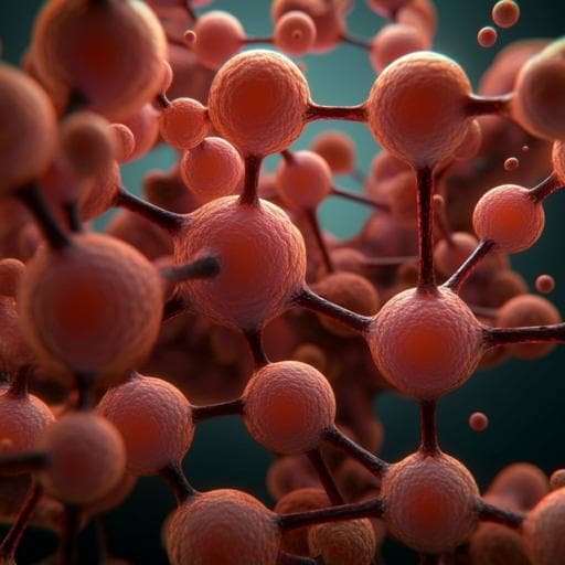
GlomSpheres as a 3D co-culture spheroid model of the kidney glomerulus for rapid drug-screening
J. Tuffin, M. Chesor, et al.
Explore these studies to deepen your understanding
Adjacent work that informs or extends this paper's methodology and findings.
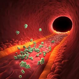
EGFP-EGF1-conjugated poly(lactic-co-glycolic acid) nanoparticles as a carrier for the delivery of CCR2– shRNA to atherosclerotic macrophage in vitro
Z. Wu, C. Chen, et al.

Evaluating the effectiveness of the Kidogo model in empowering women and strengthening their capacities to engage in paid labor opportunities through the provision of quality childcare: a study protocol for an exploratory study in Nakuru County, Kenya
K. Okelo, M. Nampijja, et al.
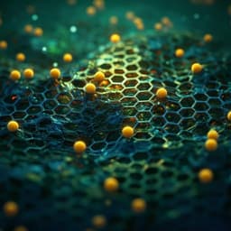
A generative artificial intelligence framework based on a molecular diffusion model for the design of metal-organic frameworks for carbon capture
H. Park, X. Yan, et al.

A cross-specific multiplicative binomial recursive model for the analysis of perinatal mortality in a diallel cross among three varieties of Iberian pig
L. Varona, J. L. Noguera, et al.


