
Medicine and HealthNature Communications
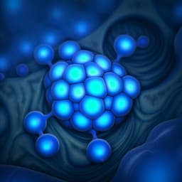
Exosome-coated oxygen nanobubble-laden hydrogel augments intracellular delivery of exosomes for enhanced wound healing
X. Han, C. Saengow, et al.
Discover a groundbreaking strategy to enhance wound healing, developed by Xiaoxue Han, Chaimongkol Saengow, Leah Ju, Wen Ren, Randy H. Ewoldt, and Joseph Irudayaraj. This innovative approach combines oxygen nanobubbles coated with exosomes from adipose tissue-derived stem cells, embedded in a self-healing hydrogel, demonstrating remarkable improvements in wound recovery.
Related Publications
Explore these studies to deepen your understanding
Adjacent work that informs or extends this paper's methodology and findings.
Medicine and Health
Development of graphitic carbon nitride quantum dots-based oxygen self-sufficient platforms for enhanced corneal crosslinking
M. Yang, T. Chen, et al.

Chemistry
The role of oxygen-vacancy in bifunctional indium oxyhydroxide catalysts for electrochemical coupling of biomass valorization with CO<sub>2</sub> conversion
F. Ye, S. Zhang, et al.
Engineering and Technology
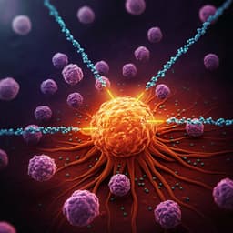
Nanoparticles and convergence of artificial intelligence for targeted drug delivery for cancer therapy: Current progress and challenges
R. P. Singh, A. Natarajan, et al.
Medicine and Health
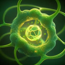
Enhanced plant-derived vesicles for nucleotide delivery for cancer therapy
S. Corvigno, Y. Liu, et al.


