Medicine and HealthNature Communications
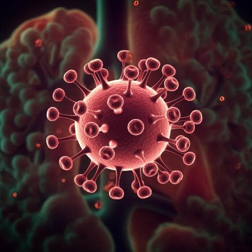
Development and evaluation of an artificial intelligence system for COVID-19 diagnosis
C. Jin, W. Chen, et al.
Discover a groundbreaking AI system developed by Cheng Jin and colleagues, designed for rapid COVID-19 detection through chest CT scans. With impressive accuracy, this deep convolutional neural network outperforms radiologists and offers speedy diagnosis, making it a revolutionary tool in medical imaging.
Related Publications
Explore these studies to deepen your understanding
Adjacent work that informs or extends this paper's methodology and findings.

Medicine and Health
An artificial intelligence system for predicting the deterioration of COVID-19 patients in the emergency department
F. E. Shamout, Y. Shen, et al.
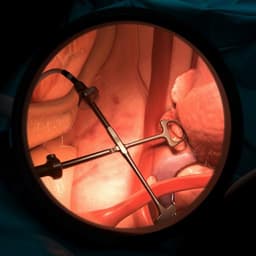
Medicine and Health
Development, deployment and scaling of operating room-ready artificial intelligence for real-time surgical decision support
S. Protserov, J. Hunter, et al.

Chemistry
ChatMOF: an artificial intelligence system for predicting and generating metal-organic frameworks using large language models
Y. Kang and J. Kim

Medicine and Health
An artificial intelligence-assisted microfluidic colorimetric wearable sensor system for monitoring of key tear biomarkers
Z. Wang, Y. Dong, et al.


