
ChemistryNature Communications
Designed Rubredoxin miniature in a fully artificial electron chain triggered by visible light
M. Chino, L. F. D. Costanzo, et al.
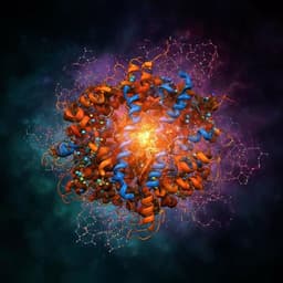
Exciting advancements in the design of metal sites in *de novo* proteins are showcased by a team of researchers including Marco Chino, Luigi Franklin Di Costanzo, and others. Their innovative 28-residue tetra-thiolate metal-binding protein exhibits remarkable precision and high reduction potential, making it a groundbreaking component in artificial light-triggered electron chains.
Related Publications
Explore these studies to deepen your understanding
Adjacent work that informs or extends this paper's methodology and findings.
Biology
Macroscopic waves, biological clocks and morphogenesis driven by light in a giant unicellular green alga
E. Afik, T. J. B. Liu, et al.
Engineering and Technology
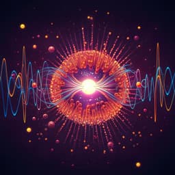
Heavy-to-light electron transition enabling real-time spectra detection of charged particles by a biocompatible semiconductor
D. Zhao, R. Gao, et al.
Chemistry
Directed ultrafast conformational changes accompany electron transfer in a photolyase as resolved by serial crystallography
A. Cellini, M. K. Shankar, et al.

Physics
Electronic transport driven by collective light-matter coupled states in a quantum device
F. Pisani, D. Gacemi, et al.


