Engineering and Technologynpj Computational Materials
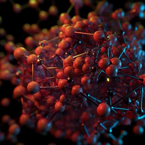
Deep learning for three-dimensional segmentation of electron microscopy images of complex ceramic materials
Y. Hirabayashi, H. Iga, et al.
This groundbreaking study by Yu Hirabayashi, Haruka Iga, Hiroki Ogawa, Shinnosuke Tokuta, Yusuke Shimada, and Akiyasu Yamamoto demonstrates the power of neural networks in recognizing intricate microstructures in polycrystalline ceramics, achieving an impressive IoU of 94.6%. Their U-Net model reconstructs giga-scale 3D images in minutes, showcasing the future of high-resolution material analysis.
Related Publications
Explore these studies to deepen your understanding
Adjacent work that informs or extends this paper's methodology and findings.

Engineering and Technology
Deep-learning-based image segmentation integrated with optical microscopy for automatically searching for two-dimensional materials
S. Masubuchi, E. Watanabe, et al.

Engineering and Technology
A generative deep learning framework for inverse design of compositionally complex bulk metallic glasses
Z. Zhou, Y. Shang, et al.
Engineering and Technology
Alternation of inverse problem approach and deep learning for lens-free microscopy image reconstruction
L. Hervé, D. C. A. Kraemer, et al.
Engineering and Technology
Rapid and flexible segmentation of electron microscopy data using few-shot machine learning
S. Akers, E. Kautz, et al.


