
Engineering and Technologynpj 2D Materials and Applications
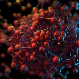
Deep-learning-based image segmentation integrated with optical microscopy for automatically searching for two-dimensional materials
S. Masubuchi, E. Watanabe, et al.
This innovative research conducted by Satoru Masubuchi and colleagues showcases a deep-learning-based image segmentation algorithm that integrates seamlessly with an autonomous robotic system, revolutionizing the automated search and cataloging of 2D materials. With the robust Mask-RCNN neural network and advanced microscopy, this technology promises to enhance efficiency in 2D material research like never before.
Related Publications
Explore these studies to deepen your understanding
Adjacent work that informs or extends this paper's methodology and findings.
Engineering and Technology
Deep learning for three-dimensional segmentation of electron microscopy images of complex ceramic materials
Y. Hirabayashi, H. Iga, et al.

Engineering and Technology
Hybrid architecture based on two-dimensional memristor crossbar array and CMOS integrated circuit for edge computing
P. Kumar, K. Zhu, et al.

Computer Science
Rewritable two-dimensional DNA-based data storage with machine learning reconstruction
C. Pan, S. K. Tabatabaei, et al.
Medicine and Health
Liquid-metal-based three-dimensional microelectrode arrays integrated with implantable ultrathin retinal prosthesis for vision restoration
W. G. Chung, J. Jang, et al.


