
Medicine and HealthNot specified in provided text
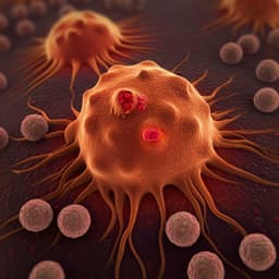
Deep Learning Approach for Early Stage Lung Cancer Detection
S. Abunajm, N. Elsayed, et al.
This groundbreaking research by Saleh Abunajm, Nelly Elsayed, Zag Elsayed, and Murat Ozer introduces a deep-learning model designed to revolutionize early lung cancer prediction and diagnosis through advanced Computed Tomography (CT) scans, aiming for unparalleled accuracy in assisting radiologists.
Related Publications
Explore these studies to deepen your understanding
Adjacent work that informs or extends this paper's methodology and findings.
Medicine and Health
Automated System for Colon Cancer Detection and Segmentation Based on Deep Learning Techniques
A. T. Azar, M. Tounsi, et al.

Medicine and Health
A multimodal deep learning approach for the prediction of cognitive decline and its effectiveness in clinical trials for Alzheimer’s disease
C. Wang, H. Tachimori, et al.

Earth Sciences
Deep multi-task learning for early warnings of dust events implemented for the Middle East
R. Sarafian, D. Nissenbaum, et al.

Education
Interpretable early warning recommendations in interactive learning environments: a deep-neural network approach based on learning behavior knowledge graph
X. Xia and W. Qi


