
Medicine and HealthCommunications Biology
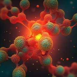
Deciphering tumour tissue organization by 3D electron microscopy and machine learning
B. D. D. Senneville, F. Z. Khoubai, et al.
This study by Baudouin Denis de Senneville, Fatma Zohra Khoubai, and their colleagues explored the intricate 3D organization of hepatoblastoma tissues. Utilizing advanced imaging techniques and machine learning, they unveiled fascinating correlations between tumor cell size and their subcellular components, advancing our understanding of tumor architecture.
Related Publications
Explore these studies to deepen your understanding
Adjacent work that informs or extends this paper's methodology and findings.

Engineering and Technology
Rapid and flexible segmentation of electron microscopy data using few-shot machine learning
S. Akers, E. Kautz, et al.

Medicine and Health
Predictive model of castration resistance in advanced prostate cancer by machine learning using genetic and clinical data: KYUCOG-1401-A study
M. Shiota, S. Nemoto, et al.
Engineering and Technology
A robust synthetic data generation framework for machine learning in high-resolution transmission electron microscopy (HRTEM)
L. R. Dacosta, K. Sytwu, et al.

Engineering and Technology
A machine learning approach to map crystal orientation by optical microscopy
M. Wittwer and M. Seita


