Medicine and Health
Concept and location neurons in the human brain provide the ‘what’ and ‘where’ in memory formation
S. Mackay, T. P. Reber, et al.
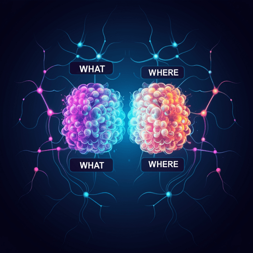
Our brains create new memories by capturing the ‘who/what’, ‘where’ and ‘when’. Measuring single-neuron activity in the human medial temporal lobe during item–location encoding, the team found two specialized groups—hippocampal, amygdala and entorhinal concept cells, and parahippocampal location-selective neurons—whose higher firing predicted successful encoding, supporting hippocampal indexing and linking the ‘what’ and ‘where’. Research conducted by Sina Mackay, Thomas P. Reber, Marcel Bausch, Jan Boström, Christian E. Elger, Florian Mormann.
~3 min • Beginner • English
Introduction
The human medial temporal lobe (MTL) is essential for memory, yet the neuronal mechanisms transforming perception into episodic memories remain largely unknown. Episodic memory integrates information about what/who occurred, where it happened, and when. Prior work in rodents, non-human primates, and humans has identified neuronal codes for these components in MTL structures. Spatial representations include hippocampal place cells and entorhinal grid cells; temporal representations such as time cells and ramping cells have also been observed. In humans, EC grid-like codes and parahippocampal involvement in spatial processing have been reported. A striking MTL finding is the existence of concept cells—neurons that respond selectively and invariantly to semantic content (e.g., familiar people, objects) across hippocampus, amygdala, and EC, closely linked to conscious perception and imagery. The PHC, in contrast, responds earlier, less selectively, and is tuned to scenes and spatial features. Previous human single-neuron studies reported memory-predictive effects mainly at the population level, with limited or inconsistent effects during encoding in selectively responsive neurons. Sparse coding may obscure detection of memory effects if only a small subset exhibits them. This study tests subsequent memory effects within visually selective neurons during encoding of item–location associations, with the goal of dissociating ‘what’ (item/concept) and ‘where’ (location) contributions to episodic memory formation.
Literature Review
- Rodent studies: hippocampal place cells and EC grid cells provide spatial maps; EC and hippocampus also show time cells and sequences reflecting event structure, suggesting a sequence-based organization of information.
- Non-human primates: EC grid-like coding of 2D space; spatial attention modulates EC receptive fields.
- Humans: fMRI/MEG show hexadirectional grid representations in EC during virtual navigation; single neurons in human EC can tune to upcoming targets; however, EC neurons may generally not process scenes/spatial information as strongly as PHC.
- PHC: linked to 3D spatial navigation and topographical learning; evidence for allocentric and egocentric representations across PHC and hippocampus; models posit hippocampal allocentric maps modulated by parietal inputs, alternating bottom-up and top-down states.
- Concept cells: selective and invariant neuronal responses to semantic concepts across hippocampus, amygdala, and EC; activated by images, text, speech, imagery, and recall; proposed as building blocks of episodic memory.
- Human single-neuron memory studies: subsequent memory effects often at population-code level (higher firing during successfully encoded or recognized items), but typically absent in selective single neurons during encoding; egocentric spatial cells are an exception. Sparse coding likely limits detectability of effects when averaging across all neurons.
Methodology
Participants: 13 in-patients (20–62 years; 8 female) with drug-resistant epilepsy undergoing invasive monitoring at University Hospital Bonn. All gave informed consent; IRB-approved.
Task design: Associative memory task on a touchscreen laptop using a 3×3 grid. During encoding, images were presented one at a time at randomly assigned grid locations. Subjects had to tap the image location during the 1.5–3.5 s presentation window to confirm perception (green outline feedback); invalid trials (missed/incorrect taps) were excluded. After encoding, a 15 s backward counting distractor (count down by 3 from 80–100) prevented rehearsal. Retrieval: each item was shown below an empty grid; subjects tapped the remembered location. Sessions lasted 35 min and consisted of multiple runs with adaptive difficulty to target ~50% accuracy by adjusting presentation duration (±0.5 s between 1.5–3.5 s) and set size (number of items per run; adjusted after three consecutive high/low performance runs). Initial set size = 2. Item pool per session: 4–8 items based on expected performance. Mean trials per session: 168.61 (SD 49.88; range 64–278); runs: 58.83 (SD 12.30; range 34–83); mean set size: 3.18 (SD 1.17; range 1–7). Retrieval and run initiation were self-paced.
Screening procedure: Morning screening identified response-eliciting images using (i) Object Screening (fixed 100 common objects/animals, 10 repeats, decision: man-made?) or (ii) Person Screening (100–150 individualized images, 6 repeats, decision: contains a face?). Images spanned many semantic concepts; selected stimuli were used in the main task.
Electrophysiology: Single-unit and multi-unit activity recorded via Behnke-Fried depth electrodes (AdTech) with 8 microwires + 1 reference per bundle, bilaterally implanted (typical scheme: 1 amygdala, 2 hippocampus, 1 EC, 1 PHC per hemisphere; up to 80 microwires). Continuous data at 32 kHz (Neuralynx ATLAS). Spike sorting with Combinato; artifact rejection included high-rate bursts, high amplitude events, overlaps, and widespread events. Manual curation classified units as single units (SU) or multi-units (MU). SNR computed (mean spike amplitude / median absolute signal); SUs had higher SNR (median 2.85) than MUs (median 2.08; P < 10^-38). Total units: 3681 (1816 SU, 1865 MU): amygdala 1117, hippocampus 1391, EC 571, PHC 602.
Responsiveness criteria: Binwise Wilcoxon rank-sum test on 100 ms windows with 50% overlap over 0–1000 ms post-stimulus, Simes correction for 19 bins, alpha = 0.001. Item responses: significant response to one/more items; location responses: significant response to one/more grid squares.
Chance-level testing: (i) Binomial test with p = 0.001 (matches alpha) per region. (ii) Permutation test with 10,000 label shuffles (items or locations) to estimate empirical sizes; p-value as proportion of shuffled proportions more extreme than observed (including half ties).
Population analyses: For each responsive neuron, averaged convolved firing rates across trials for preferred items/locations; aligned to stimulus onset (and, for PHC location cells, also to confirmation tap). Compared subsequently remembered vs. forgotten with cluster-based permutation testing (10,000 label shuffles; paired t-test threshold p < 0.05; clusters significant if top 5% of shuffled cluster sizes). Effect sizes reported as Cohen’s d.
Delay activity: For neurons responding to exactly one stimulus, computed normalized firing during 15 s counting periods; compared episodes where the preferred item’s location was later remembered vs. forgotten using paired t-tests (also per region; and for PHC location cells).
Control analyses: Re-ran main analyses excluding neurons classified as both item and location responsive; performed split-half analyses (first vs. second half of trials) to test for adaptation/interference; verified selectivity at retrieval by comparing normalized firing to preferred vs. non-preferred items/locations in the 1000 ms before the retrieval tap (signed-rank tests).
Linear mixed-effects models: Modeled relationships between subsequent memory (remembered vs. forgotten) and set size, trial duration, reaction time. For each session, computed means per subsequent memory condition; fitted LME with random intercepts and slopes for patient and session (session nested within patient). Reported fixed-effect slopes (β).
Key Findings
- Adaptive task performance correlates: Subsequently forgotten trials occurred in larger set sizes (β = −0.27, P < 10^-16) and had longer reaction times (β = 0.02 s, P = 0.01); trial duration showed no effect (β = 0.004 s, P = 0.9).
- Selective responses and counts:
• Amygdala: item neurons n = 80/1117; location neurons n = 24/1117; non-selective (both P ≥ 0.001) n = 1026/1117.
• Hippocampus: item neurons n = 81/1391; location neurons n = 26/1391; non-selective n = 1304/1391.
• Entorhinal cortex (EC): item neurons n = 32/571; location neurons n = 1/571; non-selective n = 539/571.
• Parahippocampal cortex (PHC): item neurons n = 96/602; location neurons n = 53/602 (8.80%); non-selective n = 493/602. The fraction of PHC location cells was significantly higher than in amygdala or hippocampus (chi-square x^2 = 40.50 and 53.08; both P < 10^-8). Label-shuffle empirical sizes significant for PHC location cells (0.0027), not for amygdala (0.23) or hippocampus (0.81).
- Subsequent memory effects (encoding):
• Item-selective neurons showed higher firing during subsequently remembered vs. forgotten trials in amygdala (d = 0.36, P < 10^-5), hippocampus (d = 0.33, P = 0.010), and EC (d = 0.21, P = 0.038).
• Location-selective neurons in PHC showed strong subsequent memory effects: aligned to stimulus onset d = 0.60, P < 10^-8 (1059–1444 ms window); aligned to confirmation tap d = 0.37, P < 10^-8 (25–506 ms after tap). Median confirmation latency was 1.16 s; effects were not driven by motor activity (post-tap).
• Non-selective neurons showed small effects: amygdala d = −0.10, P = 0.046 (pre-stimulus), EC d = 0.23, P = 0.031 (pre-stimulus) and d = 0.14, P = 0.005 (post-stimulus).
- Delay-period reactivation (15 s counting) showed no significant difference between later-remembered vs. forgotten associations (overall t(191) = −0.68, P = 0.50; all regions P > 0.1; PHC location-selective t(26) = −0.21, P = 0.84).
- Control analyses: Excluding neurons responsive to both item and location yielded similar effects; split-half analyses showed similar results; at retrieval, preferred stimuli elicited higher firing in all regions (all P < 0.001), and PHC location cells responded more to preferred locations (P < 0.001), supporting genuine location encoding.
Discussion
The findings dissociate ‘what’ and ‘where’ components of episodic memory encoding at the level of human single neurons in the MTL. Item-selective neurons in hippocampus, amygdala, and EC exhibited robust subsequent memory effects, consistent with the concept-cell framework in which sparse, invariant neurons act as semantic building blocks. PHC, by contrast, uniquely housed location-selective neurons that also showed strong subsequent memory effects and reactivation aligned to both stimulus onset and the confirmation tap, in line with PHC’s role in spatial processing and scene representation. Within the hippocampal indexing theory, item-selective neurons in hippocampus (and amygdala/EC) may serve as ‘pointers’ to distributed neocortical concept representations; strengthened co-activation across item-pointer neurons and PHC spatial representations via spike-timing-dependent plasticity could support binding of ‘what’ to ‘where’. The later time window for PHC location effects relative to item effects may reflect a natural hierarchy in processing narrative components (items/agents before location). EC responses resembled hippocampus/amygdala more than PHC, and showed little evidence for location coding in this map-like task, possibly because it lacked egocentric navigation demands. Overall, the results provide direct neuronal evidence that distinct selective neuron populations contribute complementary content—concepts and locations—to successful episodic encoding.
Conclusion
This study demonstrates that memory formation in humans relies on selective MTL neuron populations that differentially encode ‘what’ (item/concept cells in hippocampus, amygdala, and EC) and ‘where’ (location cells in PHC), with higher firing predicting successful subsequent recall. These results support hippocampal indexing, with concept cells functioning as pointers to neocortical semantic content, and suggest PHC location neurons as neocortical spatial representations. Future work should test whether these neurons are reactivated during consolidation (e.g., sleep), examine the integration of temporal ‘when’ information alongside ‘what/where’, and assess task- and reference-frame dependencies (egocentric vs. allocentric) for EC and PHC in more naturalistic navigation paradigms.
Limitations
- The experiment was not designed to investigate consolidation; active rehearsal was prevented with a distractor, and offline reactivation during sleep was not measured.
- Participants were patients with drug-resistant epilepsy undergoing invasive monitoring; while prior work suggests minimal interference and only small sub-populations affected during recall, generalizability to healthy populations may be limited.
- The spatial task used a constrained 2D 3×3 grid and a map-like (allocentric) format, potentially under-engaging egocentric navigation mechanisms (which might explain limited EC spatial responses).
- Sparse coding implies that effects are concentrated in small sub-populations; while addressed by pre-screening and selective analyses, undetected effects in other neuron types cannot be excluded.
Related Publications
Explore these studies to deepen your understanding of the subject.

