
Biologynature structural & molecular biology
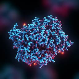
Computational design of non-porous pH-responsive antibody nanoparticles
E. C. Yang, R. Divine, et al.
Discover how a team of innovative researchers, including Erin C. Yang and Robby Divine, has designed cutting-edge octahedral nanoparticles that respond to environmental changes. These nanoparticles offer a groundbreaking method for targeted delivery of biologics, ensuring precise treatment options through antibody-mediated targeting and tunable pH-dependent disassembly.
Related Publications
Explore these studies to deepen your understanding
Adjacent work that informs or extends this paper's methodology and findings.

Biology
De novo design of pH-responsive self-assembling helical protein filaments
H. Shen, E. M. Lynch, et al.

Physics
Non-Hermitian dynamics and non-reciprocity of optically coupled nanoparticles
M. Reisenbauer, H. Rudolph, et al.

Physics
Frustrated self-assembly of non-Euclidean crystals of nanoparticles
F. Serafin, J. Lu, et al.
Medicine and Health
Computational identification of HCV neutralizing antibodies with a common HCDR3 disulfide bond motif in the antibody repertoires of infected individuals
N. G. Bozhanova, A. I. Flyak, et al.


