
Combined acid hydrolysis and fermentation improves bioactivity of citrus flavonoids in vitro and in vivo
A. König, N. Sadova, et al.
Explore these studies to deepen your understanding
Adjacent work that informs or extends this paper's methodology and findings.
Effects of swimming training in hot and cold temperatures combined with cinnamon supplementation on HbA1C levels, TBC1D1, and TBC1D4 in diabetic rats
S. M. Tayebi, A. H. Nouri, et al.
Comparative effects of high pressure processing and heat treatment on in vitro digestibility of pea protein and starch
A. E. Hall and C. I. Moraru

In vitro shear bond strength over zirconia and titanium alloy and degree of conversion of extraoral compared to intraoral self-adhesive resin cements
V. Fouquet, C. Dantagnan, et al.
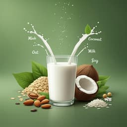
How animal milk and plant-based alternatives diverge in terms of fatty acid, amino acid, and mineral composition
S. S. Moore, A. Costa, et al.


