
PsychologyNature
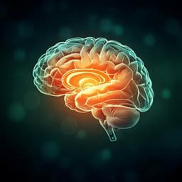
Brain-wide dynamics linking sensation to action during decision-making
A. Khilkevich, M. Lohse, et al.
Discover how brain areas collaborate during perceptual decision-making! This exciting research by Andrei Khilkevich and team reveals how sensory input and motor planning are integrated across multiple regions in the brain, leading to improved action preparation and decision-making processes.
Related Publications
Explore these studies to deepen your understanding
Adjacent work that informs or extends this paper's methodology and findings.
Medicine and Health
Event-related brain response to visual cues in individuals with Internet gaming disorder: relevance to attentional bias and decision-making
B. Kim, J. Lee, et al.

Psychology
Latent brain state dynamics distinguish behavioral variability, impaired decision-making, and inattention
W. Cai, S. L. Warren, et al.

Computer Science
Wearable EEG electronics for a Brain–AI Closed-Loop System to enhance autonomous machine decision-making
J. H. Shin, J. Kwon, et al.
Medicine and Health
Electrophysiological population dynamics reveal context dependencies during decision making in human frontal cortex
W. Shih, H. Yu, et al.


