logo
ResearchBunny Logo
Brain charts for the human lifespan

Medicine and Health

Brain charts for the human lifespan

A. 1 and A. 2

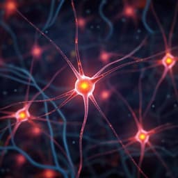
This groundbreaking research conducted by Author 1 and Author 2 introduces 'brain charts,' an interactive open-access resource utilizing MRI data to benchmark brain morphology throughout the human lifespan. By analyzing 123,984 MRI scans, the study reveals new neurodevelopmental milestones and insights into brain variation across various neurological and psychiatric disorders.

00:00
00:00
Playback language: English
Introduction
The human brain undergoes complex maturational and senescent changes throughout life. While growth charts exist for anthropometric measures like height and weight, comparable standards for brain structure are lacking. This absence hinders research on psychiatric disorders, neurodegenerative diseases, and the long-term effects of preterm birth and neurogenetic disorders. The challenges of integrating diverse neuroimaging data across studies—methodological heterogeneity, scanner differences, data processing variations—have hampered the creation of such standards. This research leverages large-scale datasets, advanced neuroimaging data processing, and statistical frameworks to overcome these obstacles and develop brain charts spanning the entire human lifespan, from mid-gestation to 100 years of age.
Literature Review
Existing literature highlights the prolonged and complex development of the brain from pregnancy to adulthood, followed by senescence. Studies emphasize the atypical brain development associated with psychiatric disorders and neurodegenerative diseases. The impact of preterm birth and neurogenetic disorders on brain structure and mental health is also documented. However, a standardized assessment tool for brain development and aging has been missing, hindering comparative analyses across studies and conditions. Previous research has explored inferring brain age from MRI data, but a comprehensive approach providing normative trajectories across the entire lifespan and accounting for technical and methodological variations in data acquisition has been lacking.
Methodology
This study aggregated MRI data from over 100 primary studies encompassing 123,984 scans from 101,457 individuals aged 115 days post-conception to 100 years. Generalized additive models for location, scale, and shape (GAMLSS) were used to model non-linear growth trajectories, stratified by sex and accounting for study-specific batch effects. The GAMLSS framework models the median, variance and skewness of the data, allowing for a robust representation of the non-linear changes in brain structure across the lifespan. Four main cerebrum tissue volumes (grey matter, white matter, subcortical grey matter, and ventricular cerebrospinal fluid) were initially analyzed. The methodology was extended to include additional MRI phenotypes, such as total surface area, mean cortical thickness, and regional volumes in 34 cortical areas. Image quality control was implemented using a combination of expert visual curation and automated metrics. Individualized centile scores were computed by benchmarking each individual scan against the normative age-related trends. A centile Mahalanobis distance (CMD) metric was developed to summarize the aggregate atypicality of an individual scan across all global MRI phenotypes. Longitudinal stability was assessed using the interquartile range (IQR) of centile scores from repeated scans. Out-of-sample centile scoring was evaluated using maximum likelihood estimation to account for study-specific offsets.
Key Findings
The study identified normative, sex-stratified trajectories for multiple brain MRI phenotypes across the lifespan. Several key neurodevelopmental milestones were identified, including peaks in grey matter volume (GMV) at 5.9 years, white matter volume (WMV) at 28.7 years, and subcortical grey matter volume at 14.4 years. The rate of growth peaked earlier for GMV and subcortical GMV (in infancy) than for WMV (in early childhood). An early period of GMV:WMV differentiation was observed, starting in the first month after birth and ending around age 3. Regional variability in volumetric neurodevelopmental trajectories was found, with primary sensory regions peaking earlier than fronto-temporal association areas. Centile scores showed significant case-control differences across various neurological and psychiatric disorders. Alzheimer's disease exhibited the largest overall difference, with the greatest effect localized to grey matter volume in females. The CMD metric ranked schizophrenia third overall in atypicality, behind Alzheimer's disease and mild cognitive impairment. Centile scores exhibited increased heritability compared to raw volumetric data. Longitudinal analyses demonstrated high stability of centile scores over time, except in individuals transitioning from mild cognitive impairment to Alzheimer's disease, where a slight increase in within-subject variability was observed. Out-of-sample centile scoring proved reliable and robust in studies with over 100 scans.
Discussion
The findings address the research question by providing normative brain charts across the lifespan. The significance lies in establishing standardized measures to quantify individual variation in brain structure. The results are relevant to the field by enabling robust comparisons across studies and diagnoses. The increased heritability of centile scores facilitates imaging-genetics studies. The ability to reliably score out-of-sample data expands the applicability of these brain charts to new research and potential clinical applications. The identification of neurodevelopmental milestones and the regional variability in growth trajectories provides valuable insights into brain development and maturation.
Conclusion
This study successfully constructed brain charts spanning the human lifespan using a large-scale aggregated MRI dataset. The charts provide standardized, age- and sex-normalized metrics to quantify typical and atypical brain development and aging. Future research should focus on expanding the brain charts to include additional MRI phenotypes, improving population representation, and validating clinical diagnostic utility. The open-access nature of the brain charts enables broad usage by the neuroimaging community.
Limitations
The dataset, although large, shows biases towards European and North American populations, limiting generalizability to other ethnicities. Data were not equally distributed across all ages, with some periods under-represented. While the statistical modeling mitigated study-specific effects, it couldn't fully correct for all limitations in primary study design, like ascertainment bias. The use of sex-stratified models followed standard practice but warrants further consideration of sex and gender.
Listen, Learn & Level Up
Over 10,000 hours of research content in 25+ fields, available in 12+ languages.
No more digging through PDFs, just hit play and absorb the world's latest research in your language, on your time.
listen to research audio papers with researchbunny