Medicine and Healthnpj Digital Medicine
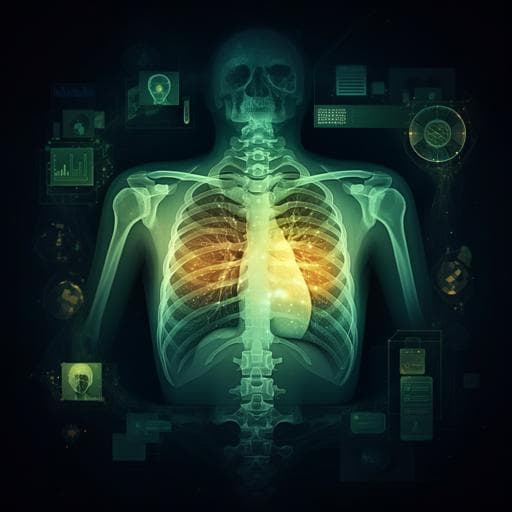
An artificial intelligence system for predicting the deterioration of COVID-19 patients in the emergency department
F. E. Shamout, Y. Shen, et al.
This research presents an innovative AI system that predicts the deterioration of COVID-19 patients in emergency settings. Leveraging a deep neural network analyzing chest X-rays alongside a gradient boosting model focused on clinical data, this system has shown considerable promise in enhancing patient triage. Conducted by a team of experts, including Farah E. Shamout and colleagues, the study emphasizes the potential of technology in improving clinical outcomes during critical times.
Related Publications
Explore these studies to deepen your understanding
Adjacent work that informs or extends this paper's methodology and findings.
Medicine and Health
Development and evaluation of an artificial intelligence system for COVID-19 diagnosis
C. Jin, W. Chen, et al.

Health and Fitness
Mobilization of expert knowledge and advice for the management of the Covid-19 emergency in Italy in 2020
S. Camporesi, F. Angeli, et al.

Psychology
The Emotional Landscape of Pregnancy and Postpartum during the COVID-19 Pandemic in Italy: A Mixed-Method Analysis Using Artificial Intelligence
Ravald, Mosconi, et al.

Psychology
An energizing role for motivation in information-seeking during the early phase of the COVID-19 pandemic
Y. Abir, C. B. Marvin, et al.


