Medicine and HealthNature Communications
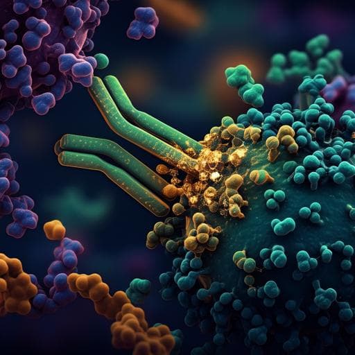
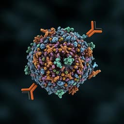
A therapeutic neutralizing antibody targeting receptor binding domain of SARS-CoV-2 spike protein
C. Kim, D. Ryu, et al.
Discover how the CT-P59 monoclonal antibody exhibits strong neutralization against SARS-CoV-2, including the D614G variant, with promising therapeutic effects demonstrated in animal models. This groundbreaking research was conducted by Cheolmin Kim, Dong-Kyun Ryu, Jihun Lee, and fellow authors.
Related Publications
Explore these studies to deepen your understanding
Adjacent work that informs or extends this paper's methodology and findings.
Medicine and Health
A variant-proof SARS-CoV-2 vaccine targeting HR1 domain in S2 subunit of spike protein
W. Pang, Y. Lu, et al.
Biology
Does the SARS-CoV-2 Spike Receptor-Binding Domain Hamper the Amyloid Transformation of Alpha-Synuclein after All?
Y. Stroylova, A. Konstantinova, et al.
Medicine and Health
Emergence and spread of a SARS-CoV-2 lineage A variant (A.23.1) with altered spike protein in Uganda
D. L. Bugembe, M. V. T. Phan, et al.
Medicine and Health
A broadly neutralizing humanized ACE2-targeting antibody against SARS-CoV-2 variants
Y. Du, R. Shi, et al.


