Engineering and Technologynpj Computational Materials
A convolutional neural network for defect classification in Bragg coherent X-ray diffraction
B. Lim, E. Bellec, et al.
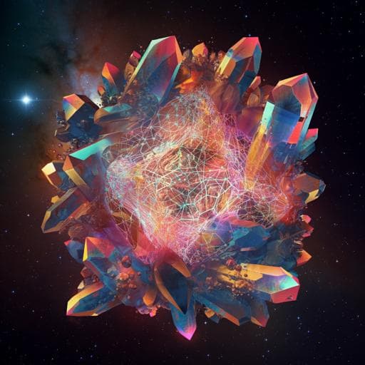
This paper highlights a groundbreaking 3D convolutional neural network (CNN) designed for swift and precise identification of defects in nanocrystals by analyzing Bragg coherent X-ray diffraction patterns. With training on extensive simulations, the CNN adeptly identifies dislocation types, marking a significant leap towards automated defect detection in materials science. This innovative research was conducted by Bruce Lim, Ewen Bellec, Maxime Dupraz, Steven Leake, Andrea Resta, Alessandro Coati, Michael Sprung, Ehud Almog, Eugen Rabkin, Tobias Schülli, and Marie-Ingrid Richard.
Related Publications
Explore these studies to deepen your understanding
Adjacent work that informs or extends this paper's methodology and findings.

Physics
Three-dimensional coherent X-ray diffraction imaging via deep convolutional neural networks
L. Wu, S. Yoo, et al.
Medicine and Health
Effectiveness of transfer learning for enhancing tumor classification with a convolutional neural network on frozen sections
Y. Kim, S. Kim, et al.
Chemistry
A deep convolutional neural network for real-time full profile analysis of big powder diffraction data
H. Dong, K. T. Butler, et al.
Psychology
Neural Representation of Multiple Languages in Polyglots: fMRI Evidence for a Shared Language Network
Malik-moraleda, Jouravlev, et al.


