Medicine and HealthNature Communications
A clinically applicable deep-learning model for detecting intracranial aneurysm in computed tomography angiography images
Z. Shi, C. Miao, et al.
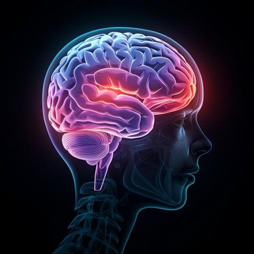
A groundbreaking study by Zhao Shi and colleagues demonstrates a deep-learning model that significantly enhances the diagnosis of intracranial aneurysms using computed tomography angiography, surpassing traditional readings by radiologists and expert neurosurgeons. This model, tested across varied imaging conditions, shows a remarkable 99.0% accuracy in predicted-negative cases, potentially reducing clinician workloads and improving patient care.
Related Publications
Explore these studies to deepen your understanding
Adjacent work that informs or extends this paper's methodology and findings.

Education
DeepLMS: a deep learning predictive model for supporting online learning in the Covid-19 era
S. B. Dias, S. J. Hadjileontiadou, et al.

Engineering and Technology
Detecting lithium plating dynamics in a solid-state battery with operando X-ray computed tomography using machine learning
Y. Huang, D. Perlmutter, et al.

Medicine and Health
A multimodal deep learning approach for the prediction of cognitive decline and its effectiveness in clinical trials for Alzheimer’s disease
C. Wang, H. Tachimori, et al.

Computer Science
Intuitive physics learning in a deep-learning model inspired by developmental psychology
L. S. Piloto, A. Weinstein, et al.


