Medicine and HealthLight: Science & Applications
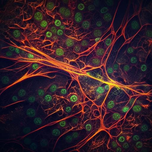
High-contrast, fast chemical imaging by coherent Raman scattering using a self-synchronized two-colour fibre laser
C. Kong, C. Pilger, et al.
This groundbreaking research by Cihang Kong and colleagues introduces a high-power self-synchronized two-colour pulsed fibre laser that enhances the capabilities of coherent Raman scattering microscopy. The improvements in imaging quality and stability allow for high-contrast imaging of living cells and tissues, expanding the possibilities for biomedical applications.
Related Publications
Explore these studies to deepen your understanding
Adjacent work that informs or extends this paper's methodology and findings.

Chemistry
Plasmon-driven chemical transformation of a secondary amide probed by surface enhanced Raman scattering
A. Dutta, M. Ončák, et al.

Health and Fitness
A tale of two paths to vaccine acceptance: self-interest and collective interest effect, mediated by institutional trust, and moderated by gender
O. Kol, D. Zimand-sheiner, et al.

Engineering and Technology
High frequency beam oscillation keyhole dynamics in laser melting revealed by in-situ x-ray imaging
Z. Wu, G. Tang, et al.

Engineering and Technology
SRS-Net: a universal framework for solving stimulated Raman scattering in nonlinear fiber-optic systems by physics-informed deep learning
Y. Song, M. Zhang, et al.


